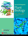
Cover Caption:
The complex of HTA and trypsin (left top). Model of the HTA-b monomer (top) and trypsin, residues 18–22 of HTA-b shown by a ball and stick model (bottom). Details of the interaction (right below). A Connolly surface diagram represents the active binding pocket of trypsin; a stick model represents only residues 18–22 of HTAb. See pages 971–982 for more details.


 Scan the QR code to view JIPB on WeChat